
- What is virus?- An infectious, obligate intracellular parasite comprising genetic material(DNA or RNA) surrounded by a protein coat and/or a membrane.‘ virus ’ (Latin, poison )
- Infectious?
- Obligate parasite?- All viruses must make mRNA that can be translated by host ribosomes; they all are parasites of host protein synthesis machinery.
- Intracellular?
- Genetic material?-RNA or DNA, single-stranded or double stranded, linear or circular
- Virus particles are designed for protection and delivery of the genome.
- Virus particles contain nonstructural components, including enzymes, small RNAs, and cellular macromolecules.
- Are viruses alive? (Yes/No/They are something in between)
- Virion: Virion(extracellular non-living but infectious particle);
- Within infected cell- Living phage
- The typical virus life cycle consists of five steps: attachment to the host cell, entry into the host cell, synthesis of viral nucleic acid and proteins within the host cell, self-assembly of virions within the host cell, and release of virions from the host cell.
- Size of virus? Very smal
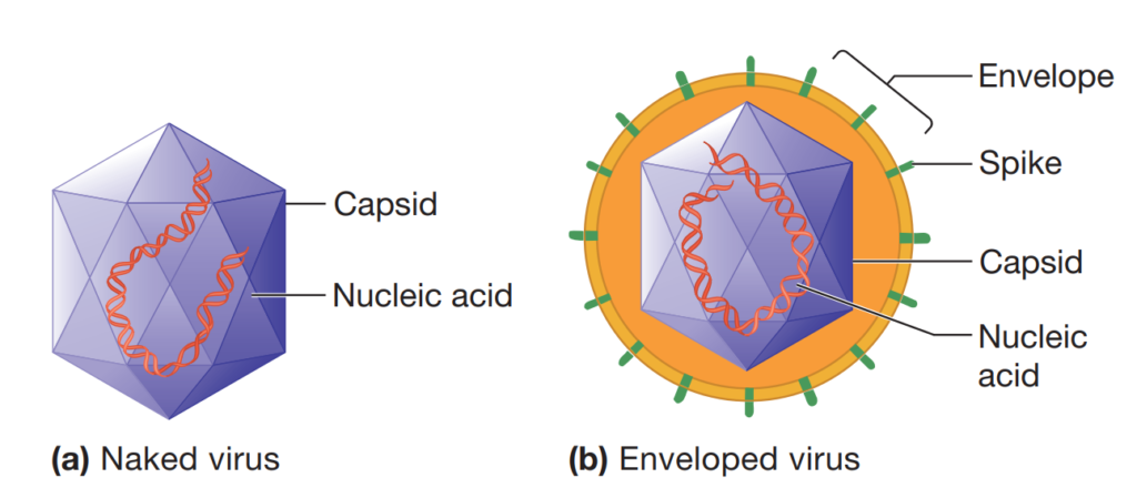
Virus= Nucleic acid+ Capsid(protein) ± Envelope(lipid)
Nucleic acid:👉Virus genome – Baltimore classification
Capsid: The capsid (from the Latin capsa for box) is the protein shell surrounding the nucleic acid genome. Capsids are large macromolecular structures that self-assemble from many copies of one or a few types of proteins. The proteins used to build the capsid are called protomers.The capsids are constructed from ring- or knob-shaped units called capsomers, each usually made of five or six protomers. Pentamers (pentons) have five subunits; hexamers (hexons) possess six. Pentamers are usually at the vertices of the icosahedron, whereas hexamers generally form its edges and triangular faces.
Amino acid–>Protomer–>Capsomers(Penton/Hexon)–>Capsid
Nucleocapsid: an unit of viral structure, consisting of a capsid with the enclosed nucleic acid.
Capsid Symmetry:
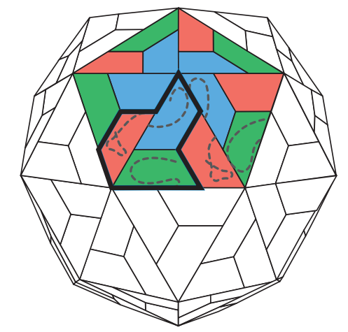
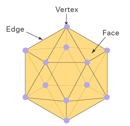
Icosahedral symmetry: Icosahedral symmetry refers to a type of symmetry seen in structures that are based on the geometry of an icosahedron, a polyhedron with 20 equilateral triangle faces, 12 vertices(corners), and 30 edges. In this symmetry, the structure can be divided into identical parts that are arranged in a way that exhibits rotational symmetry around multiple axes. As an simplest icosahedron has 20 faces, 60 identical subunits (3 per face 20 faces) is the minimal number needed to build a capsid with icosahedral symmetry. It is 5:3:2 symmetry
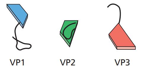
Subunit: VP1,VP2 & VP3
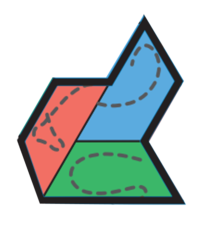
Structural unit (protomer,asymmetric unit)- Unit from which capsids or nucleocapsid are built; one or more subunits
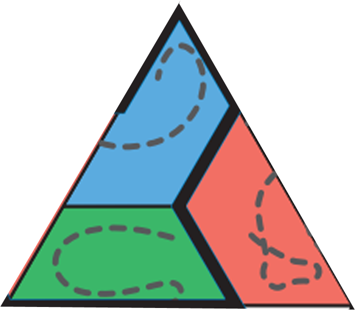
Faces(Equilateral triangle)
Capsid (capsa= Latin,box)-Protein shell surrounding genome
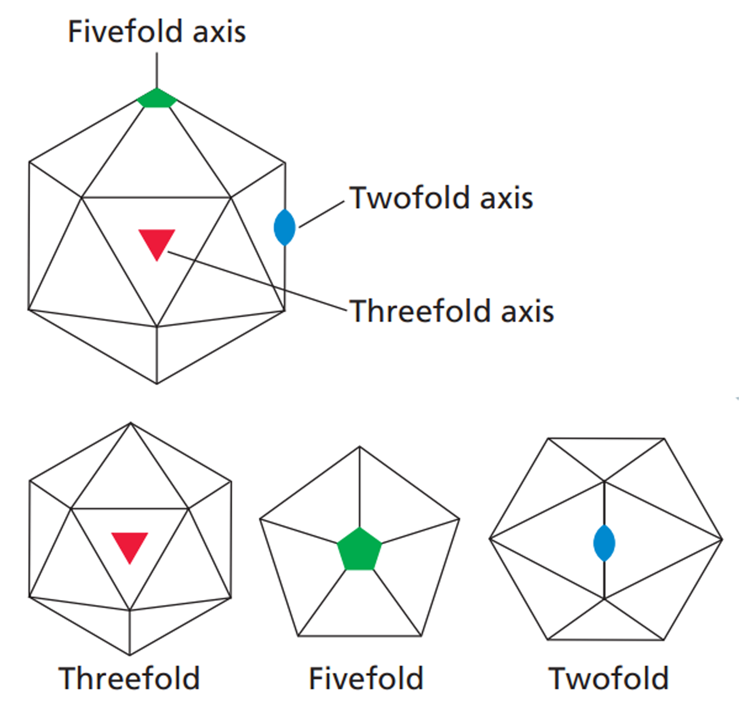
It is 5:3:2 symmetry
- Vertices shows 5 fold symmetry
- Each face shows 3 fold symmetry
- Each edge shows 2 fold symmetry
This simplest icosahedron can contain about 3kb of ssDNA/ssRNA. But this simplest 60 subunit structure restricts the genome holding capacity. Only few viruses are able to fit their genetic materials into a 60 subunit icosahedron. Most viruses require more space inside the capsid structure to hold their genome. Their capsid have more than 60 subunits. In 1962, Donald Caspar and Aaron Klug developed a theoretical framework accounting for the structural properties of larger particles with icosahedral symmetry, when a capsid contains >60 subunits, each subunit occupies a quasiequivalent position. These icosahedrons are made not just of pentamers but also of hexamers. Pentamers are arranges at vertices and hexamers at other places. Total number of subunit can be 180,240 or 780.
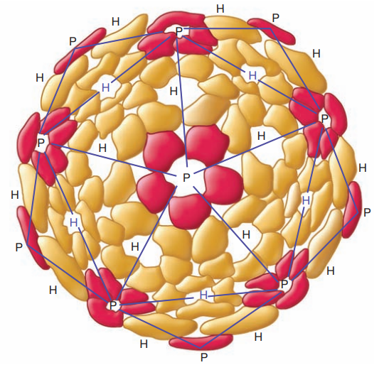
The Structure of an Icosahedral Capsid
Formed from a Single Type of Protomer. The protomers associate to form either pentons (P), shown in red, or hexons (H), shown in gold. The blue lines define the triangular faces of the icosahedron. Notice that pentons are located at the vertices and that the hexons form the edges and faces of the icosahedron. This capsid contains 42 capsomers.
Exapmle: Turnip Yellow Mosaic Virus
- Subunit= 180
- Capsomere= 32 (12 Pentamer + 20 Hexamer)
- 12 Pentamer= 12×5=60 subunit arranges at 12 verteces
- 20 Hexamer= 20×6=120 subunits arranged elsewhere
Exapmle: Adenovirus
- Subunit= 1500
- Capsomere= 252 (12 Pentamer + 240 Hexamer)
- 12 Pentamer= 12×5=60 subunit arranges at 12 vertices
- 12 long projections, fibers from vertices
- 240 Hexamer= 240×6=1440 subunits arranged faces
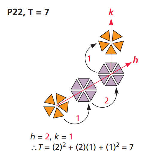
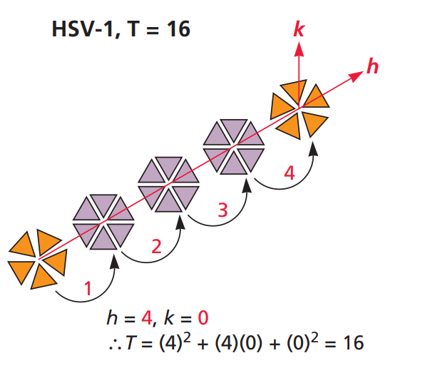
Trinagulation Number: The triangulation number, T, is the number of asymmetric units per face of the icosahedron constructed in this way. It can be shown, for example by geometry, that T = h2 + hk + k2
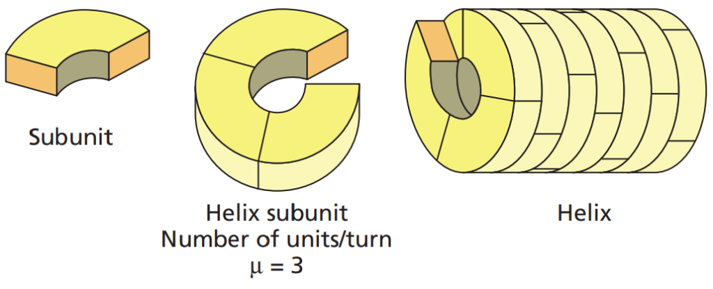
Helical symmetry: is defined by the number of structural units per helical turn, the axial rise per unit , and the pitch of the helix. Helical capsids are shaped like hollow tubes with protein walls. A key feature of a helical structure is that it can enclose any volume simply by adjusting the helix’s length. This type of structure is considered open. Few helical capsid are rigid(TMV) and few flexible; the influenza virus
genome is enclosed in thin, flexible helical capsids that are folded within an envelope. In contrast, capsids exhibiting icosahedral symmetry are closed structures with a fixed internal volume.The size of a helical capsid is determined by both its protomers and the nucleic acid it encases. The capsid’s diameter depends on the size, shape, and interactions of the protomers. The length of the capsid seems to be governed by the nucleic acid, as the capsid typically does not extend significantly beyond the end of the DNA or RNA.
Example: Vesicular stomatitis virus
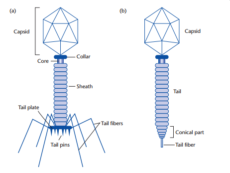
Complex Symmetry/ Binal Symmetry: While most viruses have either icosahedral or helical capsids, there are many viruses that do not fall into either category. Two notable examples are poxviruses and large bacteriophages. The T2, T4 and T6 phages (T-even phages) that infect Escherichia coli are said to have binal symmetry because they have a head that resembles an icosahedron and a tail that is
helical.Fig(a)-Bacteriophage & Fig(b)-Lambda phage
Viral Envelope Components: Some virus particles have an envelope made up of a membrane containing viral proteins, which is derived from the host cell. Envelopes are more common in animal viruses, less common in plant viruses and extremely rare in bacteriophages. These envelopes can vary greatly in size, shape, and complexity. All animal viruses share a common feature in that their envelopes are built from a lipid membrane obtained from the host cell during the viral assembly process. Animal virus envelopes usually arise from host cell nuclear or plasma membranes(cell membrane, golgi-apparatus, ER); their lipids and carbohydrates are normal host constituents. In contrast, envelope proteins are coded for by virus genes and may even project from the envelope surface as spikes, which are also called peplomers.Some spikes possess the enzyme neuraminidase, which functions in the release of mature virions from the host cell. Other spikes have hemagglutinin proteins, so named because they can bind the virions to red blood cell membranes and cause the red blood cells to clump together (agglutinate). This is called hemagglutination.Hemagglutinins play a role in the attachment of virions to host cells. Proteins, such as the spike proteins found on the outer envelope surface, are typically glycoproteins, meaning they have carbohydrates attached. In contrast, the nonglycosylated M or matrix protein is located on the inner surface of the envelope, where it helps stabilize the structure.
Virus envelope= Lipid bilayer(from host) + Viral protein(Glycoproteins)
Viral glycoproteins are integral membrane proteins firmly embedded in the lipid bilayer by a short membrane-spanning domain. These domains are having two segments: large external domain & small internal domain. External domains include binding sites for cell surface virus receptors, major antigenic determinants, and sequences that facilitate the fusion of viral and cellular membranes during entry. Internal domains, which interact with other components of the virion, are often crucial for virus assembly.
The envelopes make up the outermost layer of enveloped animal viruses. However, in bacteriophages and archaeal viruses of the PRD1 family, the membrane is located beneath an icosahedral capsid.
Enzymes:
- Neuraminidase,e.g: Influenza
- RNA dependent RNA Polymerase(that synthesizes RNA using an RNA template),e.g.: Influenza virus